Showing posts with label electron microscopy. Show all posts

2016-08-12

Tunable high-field magnetization in strongly exchange-coupled freestanding Co/CoO core/shell coaxial nanowires

German Salazar-Alvarez, Julian Geshev, Sebastia Agramunt-Puig, Carles Navau, Alvar Sanchez, Jordi Sort, and Josep Nogués

ACS Applied Materials and Interfaces, 2016
DOI: 10.1021/acsami.6b05588

Abstract:
The exchange bias properties of Co/CoO coaxial core/shell nanowires have been investigated with cooling and applied fields perpendicular to the wire axis. This configuration leads to unexpected exchange-bias effects. Firstly, the magnetization value at high fields is found to depend on the field-cooling conditions. This effect arises from the competition between the magnetic anisotropy and the Zeeman energies for cooling fields perpendicular to the wire axis. This allows imprinting pre-defined magnetization states to the AFM, as corroborated by micromagnetic simulations. Secondly, the system exhibits a high-field magnetic irreversibility, leading to open hysteresis loops, attributed to the AFM easy-axis reorientation during the reversal (effect similar to athermal training). A distinct way to manipulate the high-field magnetization in exchange-biased systems, beyond the archetypical effects, is thus experimentally and theoretically demonstrated.


2016-07-13

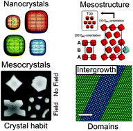
[OPEN ACCESS] Tuning the structure and habit of iron oxide mesocrystals

Erik Wetterskog*, Alice Klapper, Sabrina Disch, Elisabeth Josten, Raphaël P. Hermann, Ulrich Rücker, Thomas Brückel, Lennart Bergström and German Salazar-Alvarez*

Nanoscale (2016)
DOI10.1039/C6NR03776C


Abstract:
A precise control over the meso- and microstructure of ordered and aligned nanoparticle assemblies, i.e., mesocrystals, is essential in the quest of exploiting collective material properties for potential applications. In this work, we produce evaporation-induced self-assembled mesocrystals with different mesostructures and crystal habits based on iron oxide nanocubes by varying the nanocube size and shape, and by applying magnetic fields. A full 3D characterization of the mesocrystals was performed using image analysis, high-resolution scanning electron microscopy and Grazing Incidence Small Angle X-ray Scattering (GISAXS). This enabled structural determination of e.g. multi-domain mesocrystals with complex crystal habits, and the quantification of interparticle distances with sub-nm precision. Mesocrystals of small nanocubes (l = 8.6 – 12.6 nm) are isostructural with a body centred tetragonal (bct) lattice whereas assembly of the largest nanocubes in this study (l = 13.6 nm) additionally form a simple cubic (sc) lattice. The mesocrystal habit can be tuned from a square, hexagonal to star-like and pillar shapes depending on the particle size, shape, and the strength of the applied magnetic field. Finally, we outline a qualitative phase diagram of the evaporation-induced self-assembled superparamagnetic iron oxide nanocube mesocrystals based on nanocube edge length and magnetic field strength.


2016-07-09

3D visualization of iron oxidation state in FeO/Fe3O4 core-shell nanocubes from electron energy loss tomography

Pau Torruella, Raul Arenal, Francisco de la Peña*, Zineb Saghi, Lluís Yedra, Alberto Eljarrat, Lluis Lopez-Conesa, Marta Estrader*, Alberto López-Ortega, German Salazar-Alvarez, Josep Nogués Sanmiquel, Caterina Ducati, Paul A. Midgley, Francesca Peiró, and Sonia Estrade*

Nano Letters, 16 (2016) 5068-5073
DOI: 10.1021/acs.nanolett.6b01922


Abstract:
The physicochemical properties used in numerous advanced nanostructured devices are directly controlled by the oxidation states of their constituents. In this work we combine electron energy-loss spectroscopy, blind source separation, and computed tomography to reconstruct in three dimensions the distribution of Fe2+ and Fe3+ ions in a FeO/Fe3O4 core/shell cube-shaped nanoparticle with nanometric resolution. The results highlight the sharpness of the interface between both oxides and provide an average shell thickness, core volume, and average cube edge length measurements in agreement with the magnetic characterization of the sample.

2016-01-11

Thin Water Films at Multifaceted Hematite Particle Surfaces

Jean-François Boily, Merve Yeşilbaş, Munshi Md. Musleh Uddin, Lu Baiqing, Yulia Trushkina, and Germàn Salazar-Alvarez

Langmuir, 31 (2015) 13127–13137
DOI: 10.1021/acs.langmuir.5b03167

Abstract:
Mineral surfaces exposed to moist air stabilize nanometer- to micrometer-thick water films. This study resolves the nature of thin water film formation at multifaceted hematite (α-Fe2O3) nanoparticle surfaces with crystallographic faces resolved by selected area electron diffraction. Dynamic vapor adsorption (DVA) in the 0–19 Torr range at 298 K showed that these particles stabilize water films consisting of up to 4–5 monolayers. Modeling of these data predicts water loadings in terms of an “adsorption regime” (up to 16 H2O/nm2) involving direct water binding to hematite surface sites, and of a “condensation regime” (up to 34 H2O/nm2) involving water binding to hematite-bound water nanoclusters. Vibration spectroscopy identified the predominant hematite surface hydroxo groups (−OH, μ–OH, μ3–OH) through which first layer water molecules formed hydrogen bonds, as well as surface iron sites directly coordinating water molecules (i.e., as geminal η–(OH2)2 sites). Chemometric analyses of the vibration spectra also revealed a strong correspondence in the response of hematite surface hydroxo groups to DVA-derived water loadings. These findings point to a near-saturation of the hydrogen-bonding environment of surface hydroxo groups at a partial water vapor pressure of ∼8 Torr (∼40% relative humidity). Classical molecular dynamics (MD) resolved the interfacial water structures and hydrogen bonding populations at five representative crystallographic faces expressed in these nanoparticles. Simulations of single oriented slabs underscored the individual roles of all (hydro)oxo groups in donating and accepting hydrogen bonds with first layer water in the “adsorption regime”. These analyses pointed to the preponderance of hydrogen bond-donating −OH groups in the stabilization of thin water films. Contributions of μ–OH and μ3–OH groups are secondary, yet remain essential in the stabilization of thin water films. MD simulations also helped resolve crystallographic controls on water–water interactions occurring in the “condensation regime”. Water–water hydrogen bond populations are greatest on the (001) face, and decrease in importance in the order (001) > (012) ≈ (110) > (014) ≫ (100). Simulations of a single (∼5 nm × ∼ 6 nm × ∼ 6 nm) nanometric hematite particle terminated by the (001), (110), (012), and (100) faces also highlighted the key roles that sites at particle edges play in interconnecting thin water films grown along contiguous crystallographic faces. Hydroxo–water hydrogen bond populations showed that edges were the preferential loci of binding. These simulations also suggested that equilibration times for water binding at edges were slower than on crystallographic faces. In this regard, edges, and by extension roughened surfaces, are expected to play commanding roles in the stabilization of thin water films. Thus, in focusing on the properties of nanometric-thick water layers at hematite surfaces, this study revealed the nature of interactions between water and multifaced particle surfaces. Our results pave the way for furthering our understanding of mineral-thin water film interfacial structure and reactivity on a broader range of materials.


2015-01-07

[OPEN ACCESS] Origin of the Large Dispersion of Magnetic Properties in Nanostructured Oxides: FexO/Fe3O4 Nanoparticles as a Case Study


M. Estrader, A. López-Ortega, I. V. Golosovsky, S. Estradé, G. Salazar-Alvarez, Ll. López- Conesa, D. Tobia, E. Winkler, J. D. Ardisson, W.A.A. Macedo, M. Vasilakaki, K. N. Trohidou, R. D. Zysler, F. Peiró, L. Bergström, and J. Nogués

Nanoscale (2015)
DOI: 10.1039/C4NR06351A

Abstract:
The intimate relationship in transition-metal oxides between stoichiometry and physiochemical properties makes them appealing as tunable materials. These features become exacerbated when dealing with nanostructures. However, due to the complexity of nanoscale materials, establishing a distinct relationship between structure-morphology and functionalities is often complicated. In this regard, in the FexO/Fe3O4 system a largely unexplained broad dispersion of magnetic properties has been observed. Here we show, thanks to a comprehensive multi-technique approach, a clear correlation between magneto-structural properties in large (45 nm) and small (9 nm) FexO/Fe3O4 core/shell nanoparticles that can explain the spread of magnetic behaviors. The results reveal that while the FexO core in the large nanoparticles is antiferromagnetic and has bulk-like stoichiometry and unit-cell parameters, the FexO core in the small particles is highly non-stoichiometric and strained, displaying no significant antiferromagnetism. These results highlight the importance of ample characterization to fully understand the properties of nanostructured metal oxides.

2014-11-02

[OPEN ACCESS] Precise control over shape and size of iron oxide nanocrystals suitable for assembly into ordered particle arrays

Erik Wetterskog, Michael Agthe, Arnaud Mayence, Jekabs Grins, Dong Wang, Subhasis Rana, Anwar Ahniyaz, German Salazar-Alvarez and Lennart Bergström

Sci. Technol. Adv. Mater. 15 (2014) 055010
DOI:10.1088/1468-6996/15/5/055010

Abstract
Here we demonstrate how monodisperse iron oxide nanocubes and nanospheres with average sizes between 5 and 27 nm can be synthesized by thermal decomposition. The relative importance of the purity of the reactants, the ratio of oleic acid and sodium oleate, the maximum temperature, and the rate of temperature increase, on robust and reproducible size and shape-selective iron oxide nanoparticle synthesis are identified and discussed. The synthesis conditions that generate highly monodisperse iron oxide nanocubes suitable for producing large ordered arrays, or mesocrystals are described in detail.


2014-09-22

[OPEN ACCESS] Probing planar defects in nanoparticle superlattices by 3D small-angle electron diffraction tomography and real space imaging

Arnaud Mayence, Dong Wang, German Salazar-Alvarez, Peter Oleynikov and Lennart Bergström
Nanoscale, 2014, Accepted Manuscript
DOI: 10.1039/C4NR04156A

Abstract
We demonstrate how the acquisition and processing of 3D electron diffraction data can be extended to characterize structural features on the mesoscale, and show how lattice distortions in superlattices of self-assembled spherical Pd nanoparticles can be quantified using small-angle electron diffraction tomography (3D SA-EDT). Transmission electron microscopy real space imaging and 3D SA-EDT reveals a high density of stacking faults that was related to a competition between fcc and hcp arrangements during assembly. Information of the orientation of the stacking faults was used to make analogies between planar defects in the superlattices and Shockley partial dislocations in metallic systems.


2014-07-30

Oriented aggregation of lepidocrocite and impact on surface charge development

Philipp A. Kozin, German Salazar-Alvarez, and Jean-François Boily

Langmuir 30 (2014) 9017–9021


Abstract
The impact of lepidocrocite (γ-FeOOH) nanoparticle aggregation on mineral surface charge development was resolved in aqueous solutions of NaCl and NaClO4. Synthetic rod-like particles exhibiting charged edge (100) and neutrally/low-charged (010) faces self-aggregated in salt-free solutions. Aggregation was notably imaged by high-resolution transmission electron microscopy, and inferred by decreases in N2(g)-B.E.T. specific surface area from 94 m2/g to 77 m2/g after 12 months, and to 66 m2/g after 33 months storage. Potential determining (H+, OH–) ions loadings in the 4–11 pH range were unchanged only if the particles remained aggregated in NaCl but only if they were disaggregated in NaClO4. These differences, alongside molecular simulations and experimental ion loadings resolved in other studies from our group, point to important controls on background electrolyte ion identity on the aggregation and charge development in lepidocrocite. These results may apply further to other mineral surfaces of comparable surface (hydr)oxo populations.


2014-04-22

Postdoctoral fellowship in 3D electron microscopy studies of nanoparticles and nanoparticle arrays

Re. SU FV-1248-14. Last application date: 2014-05-09.


Stockholm University, a modern university with a multicultural environment, is one of the world’s top 100 higher education institutes. Here more than 60,000 students and 5,000 staff are active within science, the humanities and the social sciences. Stockholm is a cultural hub and economic centre, with many green areas and surrounded by water, making it an ideal place in which to enjoy a relaxed and exciting student life.
         MMK is a leading institution in materials synthesis and structure characterization. We have recently obtained a large grant for the project “3D Electron Microscopy for Nanostructure Research (3DEM- NATUR)” from the Knut and Alice Wallenberg Foundation. The 3DEM-NATUR project aims at developing new transmission electron microscopy methods to obtain 3D structural information for studying atomic and mesoscopic arrangements in solids, on surfaces and at interfaces. More information about the 3DEM-NATUR project can be found at http://www.mmk.su.se/page.php?pid=945.
         We are interested in studying nanoparticles and self-assembled nanoparticle arrays using various 3-dimensional transmission electron microscopy (TEM) techniques. Techniques involve the recently developed Rotation Electron Diffraction (RED) method and other EM techniques such as electron tomography, and STEMHAADF, STEMEELS and electron holography may also be applied.

Assessment criteria
The candidate must have obtained a PhD degree in chemistry, physics, materials science or other relevant field no more than three years prior the application deadline, unless there are special circumstances that should be taken into account. These might be illness, parental leave, union duties or similar. The applicant should have excellent English language skills, both oral and written communication.
         Proven experience in TEM and nanoparticle synthesis is essential. Experience in nanoparticle assembly and crystallography is desirable.

Terms of employment
The scholarship is for one year with the possibility of one-year extension. The preferred starting date is 4th of August 2014.

Stockholm University strives to be a workplace free from discrimination and offers equal opportunities to everyone.

More information
For further information about the position, please contact German Salazar-Alvarez, telephone +46-(0)8-163942 or german@mmk.su.se

Application
The application should be written in Swedish or English and contain:
    – Cover letter describing the interest and skills of the applicant
    – CV with publication list (including DOI),
    – Copy of PhD degree certificate,
    – Contact information for 2 reference persons

Welcome with your application, marked with the reference number SU FV-1248-14, no later than 9 May, 2014 by e-mail to: registrator@su.se.

Applications that are submitted electronically should be in Word (.doc/.docx) or PDF format. Please include the reference number SU FV-1248-14 also in the message line.

2013-12-17

Robust antiferromagnetic coupling in hard-soft bi-magnetic core/shell nanoparticles


Nature Communications 4 (2013) 2960.

M. Estrader, A. López-Ortega, S. Estradé, I. V. Golosovsky, G. Salazar-Alvarez, M. Vasilakaki, K. N. Trohidou, M. Varela, D. C. Stanley, M. Sinko, M. J. Pechan, D. J. Keavney, F. Peiró, S. Suriñach, M. D. Baró & J. Nogués


Abstract:
The growing miniaturization demand of magnetic devices is fuelling the recent interest in bi-magnetic nanoparticles as ultimate small components. One of the main goals has been to reproduce practical magnetic properties observed so far in layered systems. In this context, although useful effects such as exchange bias or spring magnets have been demonstrated in core/shell nanoparticles, other interesting key properties for devices remain elusive. Here we show a robust antiferromagnetic (AFM) coupling in core/shell nanoparticles which, in turn, leads to the foremost elucidation of positive exchange bias in bi-magnetic hard-soft systems and the remarkable regulation of the resonance field and amplitude. The AFM coupling in iron oxide—manganese oxide based, soft/hard and hard/soft, core/shell nanoparticles is demonstrated by magnetometry, ferromagnetic resonance and X-ray magnetic circular dichroism. Monte Carlo simulations prove the consistency of the AFM coupling. This unique coupling could give rise to more advanced applications of bi-magnetic core/shell nanoparticles.

2013-11-01

PhD Position in Analytical Transmission Electron Microscopy::Stockholm University, Sweden

Project title: Studies and characterization of interfaces in nanoscale materials using novel 3DEM techniques. 

Reference number: SU FV-3142-13 (project HT13-1).

Deadline: November 20, 2013

General information.

Stockholm University (http://www.su.se) is a modern university with a multicultural environment and is one of the world’s top 100 higher education institutes. Here more than 60,000 students and 5,000 staff are active within science, humanities and social sciences. The Department of Materials and Environmental Chemistry (MMK, http://www.mmk.su.se) is a leading institution with research spanning over Materials and Solid State Chemistry focusing on different classes of materials, from ceramics and glasses to self-assembled and porous materials. The work often encompasses synthesis, characterisation by X-ray diffraction and electron microscopy, NMR studies, and modelling with computer simulations. Environmental aspects are also an important part of the research activities, where refined natural or anthropogenic chemicals and materials are studied in relation to their impact in our global environment.

Project description.

Synthetic and naturally occurring nanostructured materials are the building blocks of nanoscience and nanotechnology. These nanostructures often exhibit novel properties as their physical dimensions become comparable to certain characteristic length scales, which situate them at the border between quantum effects and bulk properties.
Nanomaterials composed of two phases often shown interesting interfacial phenomena. This project focuses on developing element specific three-dimensional electron microscopy (3DEM) to obtain compositional and crystallographic information at the interface of such nanoscale materials and correlate it with optical and magnetic properties. The project will be carried out in close collaboration with other groups at Stockholm University and international partners in Spain, Denmark and USA.

Relevant publications:
1.   E. Wetterskog, C.W. Tai, J. Grins, L. Bergström and G. Salazar-Alvarez, ACS Nano 2013 7, 7132.
2.   G. Salazar-Alvarez, H. Lidbaum, A. López-Ortega, M. Estrader, K. Leifer, J. Sort, S. Suriñach, M. D. Baró, and J. Nogués, Journal of the American Chemical Society 2011 133, 16738.
3.   K.L. Krycka, J. A. Borchers, G. Salazar-Alvarez, A. López-Ortega, M. Estrader, S. Estradé, E. Winkler, R.D. Zysler, J. Sort, F. Peiró, Maria Dolors Baró, C.C. Kao, and J. Nogués, ACS Nano 2013 7, 921.
4.   S. Disch, E. Wetterskog, R. P. Hermann, G. Salazar-Alvarez, P. Busch, T. Brueckel, L. Bergström, S. Kamali, Nano Letters 2011, 11, 1651.

Funding: The project will be funded by the recently approved KAW project “3DEM-
NATUR” (http://www.mmk.su.se/page.php?pid=945).

More information.

About the project: Doc. Germán Salazar-Alvarez, Group leader, german@mmk.su.se (http://www.mmk.su.se/page.php?pid=155&id=1303)
About MMK: Prof. Gunnar Svensson, Head of Department, gunnar.svensson@mmk.su.se.

Starting date: the anticipated starting date is February 1, 2014, or later.

How to apply.

Interested candidates should send their application and supporting material to: registrator@su.se with reference number SU FV-3142-13 in the subject field.

Your application should contain:
  • An application on the form (can be found at www.mmk.su.se/page.php?pid=413) together with CV and documentation of study merits, where your eligibility is clearly documented (see below).
  • A "Letter of intent", describing your expectations of the PhD studies connected to the project.
  • IMPORTANT: Please combine all your documents into a single, self-contained pfd-file, including the cover letter.


Eligibility.

To be eligible for PhD studies in chemistry, an education at the undergraduate level of at least 240 credits is required (corresponding to three years of full-time studies on undergraduate level and one year on advanced level) with at least 60 credits in chemistry or physics. Those studies should include at least one specialized course or a thesis in the research subject. In order to facilitate the evaluation of merits and suitability for the PhD studies your curriculum vitae (CV) should contain information about the extent and focus of the academic studies. The quantity (as part of an academic year) and the quality mark of courses in chemistry and physics are of particular interest. Please, state titles of undergraduate theses and project works. Further information is found in the home page, www.mmk.su.se/page.php?pid=413.
A selection committee will assess the candidate´s ability to successfully complete the PhD program and invite short-listed candidates to an interview in person or via internet. The study merits are an important selection criterion. Economic support for the graduate studies is guaranteed for full time studies during the time agreed in the individual study syllabus (study plan), normally for four years of full time studies, see  www.mmk.su.se/page.php?pid=413.

2013-08-05

[OPEN ACCESS] Anomalous Magnetic Properties of Nanoparticles Arising from Defect Structures: Topotaxial Oxidation of Fe1-xO|Fe3-δO4 Core|Shell Nanocubes to Single-Phase Particles

ACS Nano, 2013

DOI: 10.1021/nn402487q

Erik Wetterskog , Cheuk-Wai Tai , Jekabs Grins , Lennart Bergström, and German Salazar-Alvarez


Abstract:

Here we demonstrate that the anomalous magnetic properties of iron oxide nanoparticles are correlated with defects in their interior. We studied the evolution of microstructure and magnetic properties of biphasic core|shell Fe1-xO|Fe3-δO4 nanoparticles synthesized by thermal decomposition during their topotaxial oxidation to single-phase nanoparticles. Geometric phase analysis of high-resolution electron microscopy images reveals a large interfacial strain at the core|shell interface and the development of anti-phase boundaries. Dark-field transmission electron microscopy and powder x-ray diffraction concur that, as the oxidation proceeds, the interfacial strain is released as the Fe1-xO core is removed, but that the anti-phase boundaries remain. The anti-phase boundaries result in anomalous magnetic behavior, i.e., a reduced saturation magnetization and exchange bias effects in single-phase nanoparticles. Our results indicate that internal defects play an important role in dictating the magnetic properties of iron oxide nanoparticles.

2013-01-10

Resolving Material-Specific Structures within Fe3O4|γ-Mn2O3 Core|Shell Nanoparticles Using Anomalous Small-Angle X-ray Scattering

ACS Nano 2013, 7, 921-931.

Kathryn L. Krycka, Julie A. Borchers, German Salazar-Alvarez, Alberto López-Ortega , Marta Estrader, Sonia Estradé, Elin Winkler, Roberto Daniel Zysler, Jordi Sort, Francesca Peiró, Maria Dolors Baró, Chi-Chang Kao and Josep Nogués

DOI: 10.1021/nn303600e


Abstract:

The material specific structure of monodispersed Fe3O4|γ-Mn2O3 core|shell nanoparticles is determined using multiple energy, anomalous, small-angle x-ray scattering (ASAXS). The contribution of each component to the total scattering profile is identified with unprecedented clarity. We show that Fe3O4|γ-Mn2O3 core|shell nanoparticles with a diameter of 8.2 nm ± 0.2 nm consist of a core with a composition near Fe3O4 surrounded by a (MnXFe1−X)3O4 shell with a graded composition, i.e., ranging from X ≈ 0.40 at the inner shell toward X ≈ 0.46 at the surface. Evaluation of the scattering contribution arising from the interference between material-specific layers additionally reveals the presence of Fe3O4cores without a coating shell. Importantly, the present analysis enhances the sensitivity of the method with regard to the chemical boundaries and internal nanoparticle morphology compared with traditional approaches. Finally, it is found that the material-specific scattering profile shapes and chemical compositions extracted by this method are independent of the original input chemical compositions used in the analysis, revealing multi-energy ASAXS as a powerful tool for determining internal nanostructured morphology even if the exact composition of the individual layers is not known a priori.

2012-09-01

[OPEN ACCESS] Hard and transparent films formed by nanocellulose-TiO2 nanoparticle hybrids

http://dx.doi.org/10.1371/journal.pone.0045828

PLoS ONE 2012, 7, e45828

Christina Schütz, Jordi Sort, Zoltán Bacsik, Vitaliy Oliynyk, Eva Pellicer, Andreas Fall, Lars Wågberg, Lars Berglund, Lennart Bergström, German Salazar-Alvarez

DOI: 10.1371/journal.pone.0045828


Abstract
The formation of hybrids of nanofibrillated cellulose and titania nanoparticles in aqueous media has been studied. Their transparency and mechanical behavior have been assessed by spectrophotometry and nanoindentation. The results show that limiting the titania nanoparticle concentration below 16 vol% yields a homogeneous hybrids with a very high Young’s modulus and hardness, of up to 44 GPa and 3.4 GPa, respectively, and an optical transmittance above 80 %. Electron microscopy shows that higher nanoparticle contents result in agglomeration and an inhomogeneous hybrid nanostructure with a concomitant reduction of hardness and optical transmittance. Infrared spectroscopy suggests that the nanostructure of the hybrids is controlled by electrostatic adsorption of the titania nanoparticles on the negatively charged nanocellulose surfaces.

2012-08-08

On the role of tannins and iron in the Bogolan or mud cloth dyeing process

http://trj.sagepub.com/content/early/2012/07/16/0040517512452955.abstract

Textile Research Journal, 2012, In press.


Mukta V Limaye, Zoltán Bacsik, Christina Schütz, Aïssata Dembelé, Mama Pléa, Linnéa Andersson, German Salazar-Alvarez, and Lennart Bergström


DOI: 10.1177/0040517512452955

We have investigated the chemistry of the Bogolan or mud cloth dyeing process, a traditional technique of coloring cotton cloths deeply rooted in Mali. Textiles produced by the traditional Bogolan process, using tannin-rich plant extract and iron-rich clay-based mud, were compared using infrared (IR) spectroscopy, scanning electron microscopy (SEM) and X-ray absorption near-edge spectroscopy (XANES) with cotton fibers that were impregnated with tannin and iron salt solutions. IR spectroscopy in both reflective mode on the cloth and cotton and in transmission mode on single fibers, together with SEM, showed that gallic and tannic acid adsorb and precipitate onto the cotton fiber surface. IR spectroscopy and comparison with tannin and iron solution-impregnated cotton showed that the black color of the traditional Bogolan cloth is dominated by the formation of iron-tannin complexes. The presence of iron in the Bogolan cloth was confirmed using XANES data, supporting the notion that iron has been transferred from the iron-rich clay-based mud to the cloth. The chemistry of Bogolan cloth is not only historically and culturally significant and of importance in textile conservation, but may also inspire future research on sustainable dyeing and processing techniques based on natural products.